Zur Startseite Zur Hauptnavigation Zum Inhalt Zur Suche Zur Sitemap
+A-
ENDE
  Menü
Institut für Biochemie
  Menü
  • Biochemie I
  • Biochemie II
    • Mitarbeiter
    • Publikationen
    • Forschung
      • AG Huber
      • AG Friedrich
      • AG Schmidt
      • AG Mosig
        • Mikrophysiologische Systeme
        • Über uns
        • Publikationen
        • EU-Förderung
    • Stellenangebote
    • Karrierechancen
  • Lehre
  • Anfahrt
  • Ausstellung
  • Hauptmenü
    • Patienten & Angehörige
    • Forschung
    • Lehre & Studium
    • Ärzte & Zuweiser
    • Kliniken & Institute
    • Uniklinikum Jena
    • Beruf & Karriere
  • Biochemie I
  • Biochemie II
    • Mitarbeiter
    • Publikationen
    • Forschung
      • AG Huber
      • AG Friedrich
      • AG Schmidt
      • AG Mosig
        • Mikrophysiologische Systeme
        • Über uns
        • Publikationen
        • EU-Förderung
    • Stellenangebote
    • Karrierechancen
  • Lehre
  • Anfahrt
  • Ausstellung
Institut für Biochemie / Biochemie II / Forschung / AG Mosig / Mikrophysiologische Systeme
Microphysiological Systems

Our Research Group develops microphysiological systems emulating human organ function in vitro.

We are focused on the investigation of mechanisms of human host-microbiota interaction, in particular on the orchestration of the immune response to commensal and pathogenic microorganisms.

We work in a interdisciplinary research area including microbiology, immunology, cell biology and bioengineering to evolve advanced microfluidic organ-on-chip platforms for studies on maintenance of tissue homeostasis, inflammatory disease mechanisms and the immune safety testing of drugs. Advanced tissue engineering strategies developed by our group aiming to recreate human organ models based on primary cells and human stem cells.The models are used in biomedical research as alternative to animal testing.
Our infrastructure includes fully equipped biosafety level 2 category laboratories for cell culture, tissue engineering and molecular biology. Our labs are equipped for cell isolation, work with microorganisms, stem cells and primary cell material. An advanced microfluidic platform for operation of for organ-on-chip culture under precisely controlled environmental conditions is available.

Lung-on-chip

The lung is the central organ for the exchange of gas in the human body. It is characterized by a large surface area in relation to its volume and comprises the distal branching of the trachea, bronchial tubes, bronchioles and alveoli. 

Increased exposure of the lungs to microbial pathogens can lead to pneumonia, lesion of the lung tissue (acute lung injury, ALI) and to a acute respiratory distress syndrome (ARDS). The major risk factor of ALI or ARDS is severe sepsis in 79% of cases, which may be of pulmonary (46%) or non-pulmonary (33%) origin. Pneumonia is the second most commonly acquired infectious disease worldwide with the highest population-based mortality rate. Murine models for bacterial and/or viral infections have several limitations, e.g. transmission of pathogens is inefficient in adult mice compared to the human situation. On the other hand, conventional human 2D mono-cell-culture models of the alveolus often fail to maintain differentiation and expression of alveolus-specific functions. 

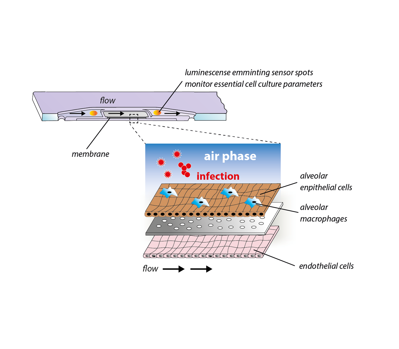
To close this gap, we have developed a immunocompetent lung-on-chip model that comprises human alveolar endothelial and epithelial cells as well as surrogates of alveolar macrophages. The model continuously produces surfactant and maintains an air-liquid-interphase for several weeks. It is able to replicate essential processes of bacterial and viral lung infections in vitro and represents a powerful platform for investigations on host-pathogen interaction and the identification of molecular and cellular targets for novel treatment strategies of pneumonia.


Gut-on-chip

In the gut commensal microorganisms of the intestinal microbiome support the digestion and absorption of nutrition by the gut. Microbial colonization is supported by the host via a mucus layer secreted by epithelial cells organized in a complex tissue comprising villi and crypts that form a tight and protective barrier between the microbiota and the circulation. A physiological communication between the members of the intestinal microbiome and their host is crucial for the maintenance of homeostasis in the human body. Thus, deregulation and imbalance of these interactions known as dysbiosis are directly associated with the development of human diseases, including diabetes, obesity, inflammatory bowel disease (IBD), cancer, depression and non-infectious inflammatory diseases caused by opportunistic pathogens.

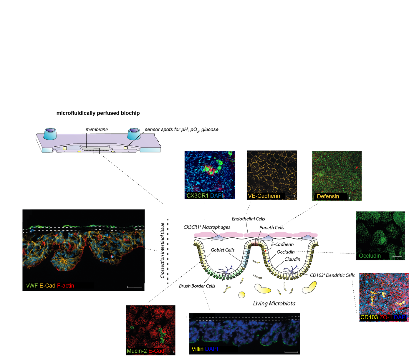
To create a platform for the investigation of the underlying mechanisms of dysbiosis associated diseases, we established a three-dimensional microphysiological model of the human intestine. This model resembles organotypic microanatomical structures and includes tissue resident innate immune cells exhibiting features of mucosal macrophages and dendritic cells. The model displays the physiological immune tolerance of the intestinal lumen to microbial-associated molecular patterns and can, therefore, be colonised with live microorganisms. It represents a valuable tool to systematically explore the underlying mechanisms of microbial communication, host-microbe interactions, microbial pathogenicity mechanisms, and immune cell activation under physiologically relevant conditions in vitro. Further, it allows the screening and development of novel treatment strategies for IBD including pharmaceutical treatments and adjustment of dysbiosis conditions to maintain physiological conditions of the human microbiota that keep opportunistic pathogens in their commensal state and prevents the onset of related inflammatory diseases.

Liver-on-chip

The liver harbors about 80% of all macrophages of the human body. Circulating monocytes are constantly patrolling within the hepatic vascular system in search of pathogen-associated molecular substances and are able to migrate into the hepatic tissue after detection of pathogens. To avoid unwanted immune responses under physiological conditions endogenous microbial molecules derived from commensal microbiota are well tolerated by the liver. However, in case of blood borne infections a prompt and robust inflammatory response is required to prevent further dissemination of the pathogen. In this context Kupffer cells, tissue resident macrophages of the liver, are central players in orchestrating a balanced immune response between immunotolerance and inflammation. 


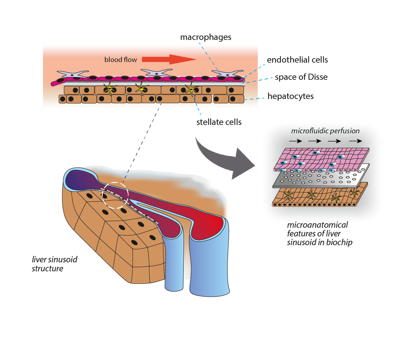
To investigate the underlying cellular and molecular mechanisms of the adapted immune reactions we developed a microfluidically perfused liver-on-chip model. The organ model includes all essential cell types of the human liver and allows the emulation of an microphysiological environment recreating organotypic functions of the liver in vitro. In the model, both inflammation-associated molecular processes of hepatocellular dysfunction as well as mechanism of organ regeneration after acute liver inflammation could be studied. The liver-on-chip platform has been successfully used in studies on drug metabolism, development of novel drug delivery systems, and disease modelling of acute and chronic liver inflammation and liver infections.

Biochemie I Ausstellung
Biochemie II Sitemap
Lehre
Anfahrt
Impressum
Datenschutz
FSU
VUD
Thüringen weltoffen
PBM
Krankenhausspiegel