Zur Startseite Zur Hauptnavigation Zum Inhalt Zum Kontakt Zur Sitemap
+A-
ENDE
  Menü
Institut für Physiologie II - Herz-Kreislauf-Physiologie
  Menü
  • Mitarbeiter
  • Forschungsthemen
    • Natriumkanäle
    • Kv-Kanäle
    • Das Selektivitätsfilter Gate von Kaliumkanälen
    • HCN-Kanäle
    • P2X-Kanäle
    • Glutamat-Rezeptoren (GPCR)
    • Lösungswechslerstrategie
    • Modellierung von Ionenkanälen
    • Acetylcholin-Rezeptoren
    • Der Spannungssensor in CNG-Kanälen
    • Rolle von CNG-Kanälen bei degenerativen Netzhauterkrankungen
    • Metabotroper Glutamat-Rezeptoren (GPCR)
  • Verbundförderungen
  • Methoden
  • Publikationen
  • Lehre
  • Kontakt
  • Karrierechancen
  • zum Institut für Physiologie I / Neurophysiologie
  • Hauptmenü
    • Patienten & Angehörige
    • Forschung
    • Lehre & Studium
    • Ärzte & Zuweiser
    • Kliniken & Institute
    • Uniklinikum Jena
    • Beruf & Karriere
  • Mitarbeiter
  • Forschungsthemen
    • Natriumkanäle
    • Kv-Kanäle
    • Das Selektivitätsfilter Gate von Kaliumkanälen
    • HCN-Kanäle
    • P2X-Kanäle
    • Glutamat-Rezeptoren (GPCR)
    • Lösungswechslerstrategie
    • Modellierung von Ionenkanälen
    • Acetylcholin-Rezeptoren
    • Der Spannungssensor in CNG-Kanälen
    • Rolle von CNG-Kanälen bei degenerativen Netzhauterkrankungen
    • Metabotroper Glutamat-Rezeptoren (GPCR)
  • Verbundförderungen
  • Methoden
  • Publikationen
  • Lehre
  • Kontakt
  • Karrierechancen
  • zum Institut für Physiologie I / Neurophysiologie
Startseite Physiologie / Forschungsthemen / Glutamat-Rezeptoren (GPCR)

Aktivierung von GPCRs mit FRET

Activation of mGluRs starts after glutamate binds into the ligand-binding domain inducing conformational changes that are transmitted through the cysteine-rich domain and end at the transmembrane domain activating the receptor and transmitting the signal inside the cell through g-proteins. Activation of mGluRs is associated with the moving of both subunits toward each other 
(Mercaggi et al. 2009, Hlavackova et al. 2012, Grushveskyi et al. 2019).
Activation of mGluRs starts after glutamate binds into the ligand-binding domain inducing conformational changes that are transmitted through the cysteine-rich domain and end at the transmembrane domain activating the receptor and transmitting the signal inside the cell through g-proteins. Activation of mGluRs is associated with the moving of both subunits toward each other (Mercaggi et al. 2009, Hlavackova et al. 2012, Grushveskyi et al. 2019).

Class C GPCRs: Metabotropic Glutamate Receptors, dimerization, and conformational dynamics

G-protein coupled receptors are the largest family of protein receptors in cell membranes, furthermore, they represent a very important group of drug targets (Krishna Sriram and Paul A. Insel, 2018). Metabotropic glutamate receptors belong to Class C of GPCRs, they are activated by glutamate, a major excitatory neurotransmitter in the brain. mGluRs are widely expressed in the CNS and play a significant role in synaptic plasticity, memory, and learning (Pin JP, Duvoisin R. (1995). They are being studied as possible drug targets for treating neurodegenerative disorders (Akito Yasuhara and Shigeyuki Chacki, 2010).

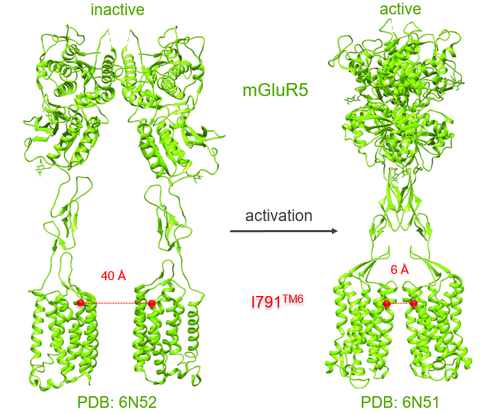
Structurally mGluRs differ from other classes of GPCRs, by forming dimers instead of monomers (Nisweder et al. 2012). The ligand-binding domain is located in the extracellular site that is followed by a long cysteine-rich domain that ends in the transmembrane region (Nisweder et al. 2010).


To monitor conformational dynamics and dimerization of mGluRs we use FRET (Föster resonance energy transfer). Fluorophores, CFP (donor), and YFP (acceptor) are fused in the intracellular loop 2 of each subunit. Conformational dynamics are monitored after applying glutamate with a fast solution exchange system in combination with a confocal laser scanning microscope. Whereas dimerization is monitored by the acceptor photobleaching method.

Methoden

FRET
Konfokale Patch-Clamp-Fluorometrie

Lösungswechslerstrategie


Themenbezogene Publikationen

Grushevskyi, E. O., Kukaj, T., Schmauder, R., Bock, A., Zabel, U., Schwabe, T., Benndorf, K., and Lohse, M. J. (2019)
Stepwise activation of a class C GPCR begins with millisecond dimer rearrangement
Proc. Natl. Acad. Sci. U S A 116, 10150-10155

Förderung

SFB Transregio ReceptorLight


Kontakt

Prof. Dr. Klaus Benndorf

Telefon: 03641 - 9 397651

Prof. Dr. Thomas Zimmer

Telefon: 03641 - 9 397655 / 9 397680
 

Dr. Ralf Schmauder

Telefon: 03641 - 9 397671

Dr. Taulant Kukaj


Telefon: 03641 - 9 397667
 

Dr. Christian Sattler

Telefon: 03641 - 9 397661
Mitarbeiter Publikationen zum Institut für Physiologie I / Neurophysiologie
Forschungsthemen Lehre Sitemap
Verbundförderungen Kontakt
Methoden Karrierechancen
Impressum
Datenschutzerklärung
FSU
VUD
Thüringen weltoffen
PBM
Krankenhausspiegel