


Pathogen-Host Interaction during Infection with Candida albicans and its Progression to Sepsis
Acronym: Candida
Principal Investigators: Dr. med. Falk A. Gonnert, Prof. Bernhard Hube
Team: Dr. Sascha Brunke, Dr. Ralf Claus, Nayla Jbeily (MSc)
Research Area: D Sepsis related Organ Failure
Project Number: D1.5
Duration: 01.08.2010 - 31.07.2015
Module: Rotational Position, Start-up Scientist



The Problem
Candida albicans dissemination via the bloodstream leads to systemic infections and sepsis. However, the early steps of attachment to and invasion into endothelia and subsequent organ colonization by C. albicans are largely unknown. This project aims to elucidate these early processes by using intravital microscopy.
Results so far
Detection of disseminating Candida albicans yeasts in live animals required the construction of effciently labelled fungal strains. Since standard GFP-labelled strains are not suitable in our system, we tagged a commonly used clinical isolate of C. albicans with the fluorescent mCherry protein for in vivo visualization. In vitro experiments showed that using the more effcient TDH3 promotor instead of the commonly used ACT1 system increased fluorescent by a factor of 10, which proved to be essential for the image acquisition in vivo. In addition, several additional fluorescent strains were created for future use (eed1∆-mCherry, cph1∆/efg1∆-mCherry, hgc1∆-mCherry, ras1∆-mCherry), all derived from previously described C. albicans mutants defective in the yeast-hyphae-transition.
An intravital microscopy model of the murine liver was adapted to allow for the observation of fungal cells within the organ. Parameters such as the age of the animals were found to severely reduce the quality of the images obtained, and hence the analysis of the colonization steps. After these improvements, fungal cells can now be observed reliably and reproducibly in the organ using our intravital microscopy system.
Starting with killed C. albicans yeasts, we observed that the temporal kinetics of fungal attachment to the liver sinusoids is significantly faster than expected. 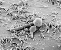 The process starts just seconds after injection of the fungi into the bloodstream, and is largely completed after only a few minutes. Especially the lateral, narrow sinusoids seem to be a preferred site of attachment. In many ways, this resembles the ad hoc adhesion to epithelial cells observed in vitro. Data acquisition is ongoing, and first steps have been taken to add a semi-automated image recognition software to aid in evaluating the time series obtained from the organ. Work planned for the near future includes investigating the early phase of yeast-hyphae-transition in vivo, and following the early immune response by additional staining of immune effector cells.
Selected publications
Brunke S, Hube B (2013) Two unlike cousins: Candida albicans and C. glabrata infection strategies. Cell Microbiol. 15(5):701-8
Lüttich A, Brunke S, Hube B, Jacobsen ID (2013) Serial passaging of Candida albicans in systemic murine infection suggests that the wild type strain SC5314 is well adapted to the murine kidney. PloS one 8: e64482
Martin R, Albrecht-Eckardt D, Brunke S, Hube B, Hünniger K, Kurzai O (2013) A Core Filamentation Response Network in Candida albicans Is Restricted to Eight Genes. PloS one 8: e58613
Mayer FL, Wilson D, Hube B (2013) Candida albicans pathogenicity mechanisms. Virulence 4: 119-128
Mayer FL, Wilson D, Hube B (2013) Hsp21 potentiates antifungal drug tolerance in Candida albicans. PloS one 8: e60417
Miramon P, Kasper L, Hube B (2013) Thriving within the host: Candida spp. interactions with phagocytic cells. Med Microbiol Immunol 202: 183-195
Pietrella D, Pandey N, Gabrielli E, Pericolini E, Perito S, Kasper L, Bistoni F, Cassone A, Hube B, Vecchiarelli A. (2013) Secreted aspartic proteases of Candida albicans activate the NLRP3 inflammasome. Eur J Immunol. 43: 679-692
Citiulo F, Jacobsen ID, Miramon P, Schild L, Brunke S, Zipfel P, Brock M, Hube B, Wilson D (2012) Candida albicans scavenges host zinc via Pra1 during endothelial invasion. PLoS Pathog 8: e1002777
Gow NA, Hube B (2012) Importance of the Candida albicans cell wall during commensalism and infection. Current Opinion in Microbiology 15: 406-412
Jacobsen ID, Wilson D, Wächtler B, Brunke S, Naglik JR, Hube B (2012) Candida albicans dimorphism as a therapeutic target. Expert Rev Anti Infect Ther. 10(1):85-93
Lüttich A, Brunke S, Hube B. (2012) Isolation and amplification of fungal RNA for microarray analysis from host samples. Methods Mol Biol. 845: 411-21
Mayer FL, Wilson D, Jacobsen ID, Miramon P, Slesiona S, Bohovych IM, Brown AJ, Hube B (2012) Small but crucial: the novel small heat shock protein Hsp21 mediates stress adaptation and virulence in Candida albicans. PloS one 7: e38584
Miramón P, Dunker C, Windecker H, Bohovych IM, Brown AJ, Kurzai O, Hube B. (2012) Cellular Responses of Candida albicans to Phagocytosis and the Extracellular Activities of Neutrophils Are Critical to Counteract Carbohydrate Starvation, Oxidative and Nitrosative Stress. PLoS One 7(12):e52850.
Contact
Tel. + 49 (0)3641 - 532 1401 oder 00
Abteilung Mikrobielle Pathogenitätsmechanismen
Leibniz Institut für Naturstoff-Forschung und Infektionsbiologie e.V.
Hans-Knöll-Institut (HKI)
Beutenbergstraße 11a
07745 Jena
Website of the Department of Microbial Pathogenicity Mechanisms (MPM) at the HKI