Zur Startseite Zur Hauptnavigation Zum Inhalt Zur Sitemap
+A-
ENDE
  menu
Klinik für Geburtsmedizin
  menu
  • We're here for you
  • Placenta-Lab
    • News
    • Team
    • Research Groups
      • CEPRE
      • AG Morales
      • AG Coxca
      • AG Schmidt, Astrid
      • AG Soczewski
      • AG Groten
      • Placenta Toxicology
    • Publications Placenta Lab
    • Alumni
  • Main menu
    • Patients & Relatives
    • Research
    • Teaching and study
    • Doctors & Referrers
    • Clinics & Institutes
    • Careers
    • University Hospital
Department of Obstetrics
  • We're here for you
  • Placenta-Lab
    • News
    • Team
    • Research Groups
      • CEPRE
      • AG Morales
      • AG Coxca
      • AG Schmidt, Astrid
      • AG Soczewski
      • AG Groten
      • Placenta Toxicology
    • Publications Placenta Lab
    • Alumni
Department of Obstetrics / Placenta-Lab / Research Groups / AG Groten
Prof. Dr. med. Tanja Groten
Direktorin der Klinik und Poliklinik für Geburtsmedizin Köln,
CEPRE SP4

Web

Members

Dr. rer. nat. Silke Große
Placenta Labor,
Post-Doc,
AG Groten

Telefon: 329293
Dr. med. Anna Multhaup
AG Groten

Theresa Forndran
cand. Dr. med.,
AG Groten
Lara Hausdorf
cand. Dr. med.,
AG Groten
Vanessa Leske
cand. Dr. med.,
AG Groten
Lena Lode
cand. Dr. med.,
AG Groten
Jasmin Schneebeck
cand. Dr. med.,
AG Groten

Gina Weber
cand. Dr. med.,
AG Groten
Emma Pusch
Bachelorstudent,
AG Groten

Alumni

Dr. med. Michael Schönleben

Dr. med. Veronika Teichert

Subject

In our research group, we focus on the role of maternal vasculature in placenta-associated pregnancy disorders such as growth restriction and preeclampsia. In addition to our clinical studies on the endothelial health of mothers with placental disorders, we are conducting experimental studies in the laboratory on endothelial cell dysfunction and the interaction of trophoblast and endothelial cells. The subject of these studies is the interaction between endothelial and placental function and dysfunction. Because clinical findings indicate that women with pre-aged endothelial health are more likely to develop placenta-associated pregnancy complications, our current projects are investigating the association of cellular aging processes in endothelial cells and in the placenta with these pregnancy complications.

Because of the close association of endothelial and placental dysfunction with diabetes mellitus, our latest project focuses on investigating the specifics of placental function in patients with diabetes during pregnancy. As part of this, we are also analyzing our clinical data collected during the care of pregnant women with diabetes at our clinic's Center of Excellence for Diabetes and Pregnancy.

Background

Fetal growth restriction and preeclampsia

Die Plazenta bildet die direkte Grenzfläche zwischen maternalem Blut und fetalem Gewebe. Die Oberfläche der Plazenta wird von speziell ausgereiften Trophoblastzellen überzogen, die ein Syncytium bilden und als Syncytiotrophoblast bezeichnet werden. Dieser Syncytiotrophoblast unterliegt während der gesamten Schwangerschaft Umbauprozessen, während derer einerseits apoptotisches Zellmaterial von der syncytialen Oberfläche abgeschnürt und an die Mutter abgeben wird und andererseits der Syncytiotrophoblast von fusionierenden Cytotrophoblasten der darunter liegenden Schicht ständig erneuert wird. Das an die Mutter abgegebene apoptotische Material ist im mütterlichen Blut in Form von plazentaren Mikropartikeln nachweisbar und kann eine maternale Endothelstörung, wie sie bei der Präeklampsie vorliegt, verursachen.

Bei Schwangerschaften mit fetalen Wachstumsrestriktion zeigt sich eine unvollständige Umwandlung der maternalen Spiralarterien in widerstandsfreie, weite Gefäße. Eine geringere Invasivität und Interaktion von Trophoblastzellen mit dem Endothel der mütterlichen Gefäße wird als pathogenetischer Mechanismus angenommen. Die mangelnde Gefäßumwandlung führt zu einer Minderperfusion der Plazenta und damit nicht nur zur Minderversorgung des Feten und zur Wachstumsrestriktion, sondern möglicherweise auch zu Funktionsstörungen des Trophoblasten. Es kommt zu einer vermehrten Abschnürung von Mikropartikeln mit Endothel-aktivierendem Potential. Zusätzlich wird der endothelschützende plazentare Wachstumsfaktor PlGF nur in geringer Menge gebildet und an die Mutter abgegeben.

Es ist nicht geklärt, ob die Endothelaktivierung bei der Mutter den plazentaren Veränderungen möglicherweise vorrausgeht. Nachgewiesen ist, dass es bei Schwangeren mit Präeklampsie und Wachstumsrestriktion zu einer Erhöhung gefäßaktivierender und von von aktivierten Gefäßen sezernierten Faktoren im Blut kommt. Bekannt ist darüber hinaus, dass diese Frauen, lebenslang ein erhöhtes Risiko haben an kardiovaskulären Erkrankungen zu leiden und auch zu sterben (Melchiorre 2012, Lancet 2002).

Unklar ist, ob diese Langzeitfolgen aufgrund der Schwangerschaftskomplikation entstehen, oder ob die Schwangerschaftskomplikation Folge einer vorbestehenden maternalen Endothelstörung sind.

Die Fragestellung einiger Projekte in unserer Arbeitsgruppe ist es deshalb die molekularen Mechanismen der endothelialen Dysfunktion bei der Präeklampsie und die Frage nach der Bedeutung der maternalen Endothelfunktion für die Entstehung der Präeklampsie und der fetalen Wachstumsrestriktion zu untersuchen.

Diabetes mellitus

Das Risiko für eine plötzliche Plazentainsuffizienz ist im Rahmen von Diabetes in der Schwangerschaft, insbesondere bei schlecht eingestelltem Diabetes mit anhaltender maternaler Hyperglykämie, deutlich erhöht und es kommt schlimmstenfalls zum intrauterinen Versterben des Kindes. Dabei bleiben die geburtshilflichen Überwachungsinstrumente oft unauffällig. Dies betrifft insbesondere auch die Messungen der feto-plazentaren Perfusion, die bei Plazentainsuffizienz, wie sie zur Wachstumsrestriktion der Kinder führt, immer auffällig werden und einen erhöhten plazentaren Widerstand bei Untergang von Plazentagewebe anzeigen. Diese Veränderungen zeigen sich bei Diabetikerinnen nicht, obwohl postpartale Blutgasanalysen übereinstimmend zeigen, dass diabetische Plazenten eine geringere Oxygenierungskapazität aufweisen. Histomorphologisch sind bisher in diabetischen Plazenten keine typischen Veränderungen, wie sie bei der Plazentainsuffizienz auftreten, beschrieben. Tatsächlich wurde jedoch in verschiedenen Arbeiten eine vermehrte Fibrose des villösen Stromas in diabetischen Plazenten. Als Fibrose wird eine Gewebeveränderung bezeichnet, die durch die pathologische Vermehrung von Bindegewebszellen und Kollagenfasern gekennzeichnet ist, die letztlich zu einer Organdysfunktion führt. Dabei wird nicht nur direkt die Matrixsynthese durch vorhandene Fibroblasten stimuliert, sondern auch die Differenzierung von Immun-, Epithel- und Endothelzellen in einen Fibroblasten-ähnlichen Phänotyp initiiert – die sogenannte Epithelial-Mesenchymale-Transition (EMT).

Hohe Glukose stimuliert fibrogene Signalwege, indem sie die Bildung reaktiver Sauerstoffspezies auslöst, Wachstumsfaktorkaskaden (wie TGF-beta/Smad3 und PDGFs) aktiviert, proinflammatorische Zytokine und Chemokine induziert. Auch in der Plazenta ist daher unter dem Einfluss erhöhter mütterlicher Glukosespiegel eine Induktion von EMT in Trophoblastzellen denkbar, aber bisher nicht gezeigt oder untersucht. EMT, bei der funktionelles Trophoblastgewebe in Bindegewebe umgewandelt würde, könnte eine Erklärung für den beobachteten Verlust der Oxygenierungskapazität der Plazenta sein.

Die Fragestellung unseres neuesten Projektes unserer Arbeitsgruppe ist es die molekularen Mechanismen der Plazentaren Dysfunktion bei Diabetes mellitus in der Schwangerschaft zu untersuchen.

Current projects

Molecular mechanisms of action of the NO donor pentaerytrithyltetranitrate (2024, MD project Vanessa Leske)

Investigation of the molecular mechanisms of the effect of the NO donor pentaerytrithyltetranitrate on the regulation of cellular stress

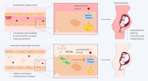
Preeclampsia and fetal growth restriction are pregnancy complications characterized by placental and endothelial dysfunction. In a clinical study, we have shown that the NO donor pentaerythrityltetranitrate (PETN) reduces the rate of pregnancy-induced hypertension in high-risk pregnancies by 30 % and simultaneously lowers the concentration of endothelial activating cytokines in the mothers' blood. Translationally, we were able to show that in stressed endothelial and trophoblast cells in the presence of PETN, the formation of reactive oxygen species and cellular dysfunction in response to oxidative stress is reduced. It is known from literature that PETN induces the expression of the antioxidant enzyme heme-oxigenase-1 (HO-1) in endothelial cells. The exact molecular mechanisms of the demonstrated effect of PETN have not yet been investigated. In this project, the signaling cascades stimulated by PETN will be identified by omic analysis and verified by Western blot. The clarification of the mechanisms of action of PETN is the basis for the identification of further indications for the use of PETN in diseases associated with endothelial dysfunction in humans.

Chronological vs. stress-induced placental aging (JSAM 2023, MD project Gina Weber).

Does the placenta age or stress itself to death? – Chronologiccal or stress-induced senescence as the predominant phenotype in the placenta

Aging of the placenta is a normal physiological phenomenon. The placenta develops, matures and ages over the course of 40 pregnancy weeks. As the pregnancy progresses, senescence markers are increasingly expressed in the placenta as well as oxidative stress accumulates. In pregnancy complications associated with placental insufficiency and oxidative stress there is an increased and earlier expression of senescence markers.

Based on these observations we ask the question whether the aging shown in the placenta is predetermined (chronological senescence) or whether it is a consequence of oxidative stress accumulated during pregnancy (stress-induced senescence). We would like to answer this question by simultaneously staining markers for senescence, oxidative stress and antioxidant capacity in the cytotrophoblast, the functional part of the placenta by multiplex immunofluorescence staining (mIF). For this purpose we will investigate FFPE (formalin fixed paraffin embedded) samples from healthy (trimester 1, 2, 3) and pathological (trimester 3) placentas collected in the placenta bank of the University Hospital Jena. In addition, we will investigate whether senescence-associated cytokine expression in explants of fresh placental samples is pathology-associated.

Fibrosis in the placenta after pregnancy in diabetes (IZKF 2023, MD project Lara Hausdorf)

Trophoblastic epithelial-mesenchymal transition with consecutive fibrosis: a cause for reduced placental function in hyperglycemic pregnancies?

Overall perinatal mortality continues to decline and is currently 0.55 % in Germany. In pregnant women with preexisting diabetes, however, perinatal mortality remains high, despite improved care, and is 1.5 to 2 % in various European studies. Often, sudden failure of placental function occurs close to term, and infants die in utero (Hug 2021). In this case, the usual obstetric monitoring tools are not effective. Histologically, the changes typical of placental insufficiency cannot be detected in these placentas either (Huynh 2014). However, increased fibrosis of these placentas has been demonstrated in a few studies (Salge 2012, Dasgupta 2022, Abdelhalim 2018). Functional studies also show, impaired oxygenation in diabetic placentas (Taricco 2009). Diabetes in pregnancy is associated with persistent maternal hyperglycemia, especially in poorly controlled diabetes, and fibrosis is known to develop in various organs under the influence of hyperglycemia. Functional epithelial tissue is replaced by connective tissue, which is associated with a loss of function. We aim to investigate whether glucose can induce epithelial-mesenchymal transition (EMT) and the development of fibrosis in vitro using trophoblast cell lines and immunohistochemistry in preparations of healthy and type 1 diabetes mellitus placentas.

Preeclampsia-associated vascular aging (JSAM 2022, MD project Lena Lode)

In preeclampsia (PE) and fetal growth restriction (FGR), endothelial dysfunction exists during pregnancy. Affected women are at increased risk for cardiovascular disease throughout life. Increased cardiovascular risk is generally associated with premature aging of the vascular system, particularly the endothelium. It is postulated that PE leads to aging of the endothelium and that a prematurely aged endothelium promotes the development of PE. Premature endothelial aging is often caused by cellular and oxidative stress. An important antioxidant and anti-inflammatory enzyme is heme oxigenase-1 (HO-1). HO-1 expression is induced in endothelial cells (EC) by the NO donor pentaerythrityl tetranitrate (PETN). To date, there is no treatment for PE; however, PETN was used in a clinical trial and was shown to improve maternal clinical outcome. In experimental studies, PETN reduced the effect of oxidative stress in EC. In this project, we will use a cell culture model to investigate whether PE simulating stimuli lead to aging of EC and whether EC aged by oxidative stress respond more strongly to these stimuli. In addition, the effect of PETN in the context of senescence induction will be investigated.

Senescence markers in the placenta (JSAM 2021, MD project Theresa Forndran)

Senescence markers in the placenta - Premature aging as a consequence of or cause of pregnancy pathologies.

The placenta develops, grows, differentiates, and ages during the 9 to 10 months of pregnancy. Physiologically, the functionality of the placenta decreases markedly after 40 weeks of gestation and it begins to fail altogether, which is clinically evident in an increased intrauterine mortality rate of human fetuses after 38 weeks of gestation.

Accelerated and premature aging of the placenta represents a pathological condition (Fabiana 2020) and is a hallmark of several placenta-associated pregnancy complications, such as fetal growth restriction (FGR) (Paules 2019) and preeclampsia (PE) (Farladansky-Gershnabel 2019)-the two leading causes of maternal and neonatal morbidity and mortality. Understanding the physiologically occurring placental aging processes and how they are disrupted is therefore of enormous clinical importance especially to develop clinical approaches to treatment.

The cellular phenotype of aging is senescence, in which irreversible cell cycle arrest occurs (Yosef 2016). In this process, the cyclin kinase inhibitors p21, p16, and p53 are increasingly expressed in the cell (Kruiswijk 2015, Childs 2014). Senescent cells produce proinflammatory cytokines called "senescence-associated secretory proteins" (SASP) that accelerate aging processes in neighboring cells (Herranz 2015, Laberge 2015). The composition of SASP varies depending on the cell, with IL-6 (interleukin-6) and TNF-α (tumor necrosis factor-alpha) as markers. At the cellular level, placental aging is seen through the expression of these senescence markers (Higuchi 2019).

There is a causal relationship between oxidative stress leading to the production of ROS (reactive oxygen species) and cell senescence (Passos 2010, Cindrova Daviesa 2018), suggesting that oxidative stress contributes to premature aging. ROS induce oxidation of polyunsaturated fatty acids, leading to increased aldehyde synthesis such as that of 4-hydroxy-2-nonenal (HNE). ROS production is counteracted by antioxidants such as superoxide dismutase (SOD) (Stadtman and Levine 2003, Panieri 2013, Forman 2008). Microarray and proteomic analyses of human placentas of different gestational ages have shown that the expression of p21, p53, and SASP, as well as HNE and SOD activity, is increased early in placentas from patients with PE and FGR (Guerby 2021).

Cell fusion, an essential physiological process for the establishment and expansion of the syncytiotrophoblast (outer cell layer of the placental barrier), has recently been identified as a trigger of cell senescence (Cox and Redman 2017, Redman 2020). The syncytiotrophoblast shows, the high expression of the cyclin kinase inhibitors p16, p21, and p53 characteristic of senescent cells, as well as the secretion of SASP, which induces labor toward the end of pregnancy (Menon 2016a, Menon 2016b, Phillippe 2015). In cases where fusion is restricted, the syncytiotrophoblast is hypofunctional. As a result, the mother may develop preeclampsia and the fetus may develop growth restriction. Premature senescence in placental membranes may result in preterm labor and delivery (Cox and Redman 2017).

The goal of this project is to immunohistochemically analyze the expression of several known senescence markers, such as p21, p16, and p53, markers of oxidative stress, and SASP, in placentas from patients with PE and/or FGR.

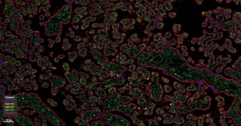
To functionally characterize the affected placental cell populations and their interactions, we will use spatial phenotyping to assign cell type and cell senescence status after multiplex immunofluorescence labeling (multiplex IF) and thus draw conclusions about their role in placental senescence in the different pregnancy pathologies.

After establishing multifluorescence-based immunodetection using control placentas from healthy pregnant women with term births, comparative evaluation with retrospectively collected placental blocks from clinically diagnosed PE and FGR pregnancies will be performed. Finally, the influence of PETN (pentaerythritol tetranitrate), a drug investigated in recent years in the PETN study for the treatment of pregnant patients at risk for developing FGR and/or PE (Groten 2019), on the expression patterns of placental senescent markers will be analyzed.

Influence of PETN on trophoblastic stress reaction (MD project Jasmin Müller)

Influence of  PETN on trophoblastic stress reaction

Completed projects

Influence of PETN on endothelial stress response (IZKF, JSMC, MD project Veronika Teichert).

PETN-induced antioxidant properties in endothelial cells as a target for secondary prevention of endothelial dysfunction in pregnancy.

These studies will lay the basic scientific foundation for clinical studies demonstrating the protective effect of PETN in pregnant women at increased risk for fetal growth restriction (FGR). Thus, this project is a typical example of translational research.

According to current knowledge, increased release of endothelium-activating soluble VEGF receptor-1 (sFlt-1) and endoglin from the placenta occurs in preeclampsia due to impaired trophoblast differentiation in early pregnancy (Huppertz 2008, Roberts 2009). At the same time, the endothelium-protective placental growth factor PlGF is produced in a reduced manner. There is an imbalance of endothelium-protective and endothelium-activating factors in the maternal circulation, leading to generalized endothelial dysfunction with coagulation activation, increase in endothelial permeability, and blood pressure elevation (Asif Ahmed and Wenda Ramma 2011). In FGR, trophoblast invasion and subsequently adequate conversion of spiral arteries into nonresistant vessels is insufficient. Perfusion of the placenta is impaired. FGR also results in impaired vascular function with increased sFlt-1 and decreased PlGF in the maternal circulation (Taylor 2003). Women who develop PE or FGR during pregnancy are at increased risk for cardiovascular disease throughout life due to impaired endothelial function (Brown et al. 2013).

NO donors cause vascular dilatation and thus a reduction in blood pressure. Continuous therapy with nitrates such as GTN leads to a decrease in the effect, the so-called nitrate tolerance. Taking the NO donor pentaerytrityl tetranitrate (PETN) does not show nitrate intolerance, but long-lasting vasodilation (Daiber et al. 2008). Furthermore, PETN induces the expression of the antioxidant heme oxygenase-1 (HO-1) in endothelial cells (Pautz et al. 2009, Daiber et al. 2012). The mechanism of action of HO-1 is based on the degradation of heme to carbon monoxide (CO), iron, and biliverdin, which is degraded to bilirubin (Sikorski et al. 2004). In this process, CO additionally acts as a vasodilator and bilirubin as an antioxidant. (George et al. 2014). In animal models, it has been shown that PETN-induced expression of HO-1 resulted in a decrease in the formation of oxygen radicals and a reduction in blood pressure without the emergence of nitrate tolerance (Schuhmacher et al. 2010, Wenzel et al. 2007). PETN furthermore slows down the progression of atherosclerosis as a consequence of increased expression of HO-1 (Polte et al. 2000, Oppermann et al. 2009). Conversely, in the absence of HO-1, there is extensive endothelial dysfunction with coagulation activation, intravascular hemolysis, and endothelial death (Yachie et al. 1999).

In a clinical trial at the University Women's Hospital in Jena, Germany, treatment with PETN was shown to reduce the risk of FGR, perinatal death, preterm delivery, and premature placental abruption in high-risk patients. In addition, there was a significantly milder course of PE that occurred later in pregnancy and was less frequently associated with preterm delivery and severe FGR (Schleussner et al. 2014). The reason for the more favorable course of the disease, in addition to the vasodilatory one, is most likely explained by the endothelium-protective effect of PETN. This relationship will be investigated and further elucidated in vitro in the present work.

HUVEC monolayers are used as a cellular model to study endothelial dysfunction in cell culture. Intact monolayer function is indicated by constant density with constant electrical resistance in impedance measurement in the XCelligence® system, continuous staining for interendothelial adhesion molecules such as VE-cadherin in immunocytology, and activation of survival signals by VEGF. These functions are disrupted by the addition of endothelial activating agents. The dysfunction of the endothelial monolayer in cell culture is then manifested by the breakdown of the barrier in the XCelligence® system, immunocytologically in a discontinuous staining pattern of VE-cadherin, in the release of endothelial activators, such as sFlt-1 into the cell culture medium and activation of signal transduction pathways mediating increased proliferation and migration of endothelial cells. In this process, signaling in endothelial cells is mediated mainly via VEGF receptor 2, which is in complex with VE-cadherin at the cell membrane in the stable endothelial cell monolayer and activates survival pathways in cells via PI3K and AKT.

In dysfunctional monolayers, activation of VEGFR2 via PKC activates ERK1/2 and stimulates migration and proliferation (Cross 2003): activation of VEGFR-2 with phosphorylation of the receptor at PY99 then also leads to the release of sFlt-1 (Cudmore 2007). It has been shown that HO-1 induction in endothelial cells leads to decreased release of sFlt-1 (Cudmore 2007). In vivo, HO-1 expression is triggered by various effectors in endothelial cells. Subsequently, it exerts intracellular antiapoptotic, antiproliferative, and anti-inflammatory effects via the accumulation of heme and CO (Loboda, 2016).

The role of adhesion molecules during trophoblast invasion and spiral artery maturation (MD project Anna Multhaup)

Studies on the role of adhesion molecules in the interaction of trophoblast cells and endothelial cells during trophoblast invasion and spiral artery maturation.

Defective trophoblast invasion during placentation results in significantly fewer trophoblast cells attached to the maternal endothelium (Kaufmann et al. 2003, Pijnenborg et al. 2006). This results in a lack of transformation of the maternal spiral arteries with subsequent reduced supply to the fetus and possible pregnancy complications (Cetin and Antonazzo 2009). The starting point of this work was the observation that disruption of differential expression of adhesion molecules on extravillous trophoblast cells was a possible cause of their reduced invasion and incomplete vascular maturation (Damsky and Fisher 1998, Zhou et al. 1997a).

Because the migration of trophoblast cells along maternal spiral arteries resembles the recruitment of neutrophil granulocytes to the endothelium during acute inflammation, we hypothesized that vascular invasion of trophoblast cells is facilitated and regulated by the expression of similar adhesion molecules (Burrows et al 1994). The aim of this work was to identify the importance of selected adhesion molecules of extravillous trophoblast cells, for their interaction with maternal endothelial cells during placentation.

For this purpose, the expression of the selected adhesion molecules was manipulated in the extravillous trophoblast cell line HTR-8/SVneo, and the resulting changes were analyzed for their interaction with primary endothelial cells HUVEC in a three-dimensional cell culture model on Matrigel®. Suppression of adhesion molecule expression was achieved by transfection of specific siRNA and monitored by protein and gene analysis. Cell interaction was analyzed microscopically and the images obtained were quantified in an objective and standardized manner using the Wimasis Image Analysis® internet platform and then statistically analyzed.

A statistically significant involvement of N-cadherin in the migration and interaction of HTR-8/SVneo with HUVEC cells in a 3D coculture on Matrigel® was demonstrated. This suggests a relevance of this molecule in trophoblast invasion in vivo. Also after suppression of the expression of CD162 a reduced interaction of the cell lines is shown, which however is not statistically significant after quantitative analysis.

We demonstrated that in the cell culture model adhesion molecules have a relevant influence on cell interaction and may play a key role in the multifactorial event of placental dysfunction.

Studies on the role of miRNA complex 371-3 in trophoblast invasion (MD project Michael Schönleben)

Role of miRNA complex 371-3 in epithelial-mesenchymal transition.

Image source:

https://smart.servier.com/ (pregnant woman)

We're here for you
Placenta-Lab