Zur Startseite Zur Hauptnavigation Zum Inhalt Zur Suche Zur Sitemap
+A-
ENDE
  menu
Institut für Humangenetik
  menu
  • Homepage
  • General Information
  • Fields of work
    • Functional Genetics
    • Molecular Genetics
    • CCB Lab
    • Molecular Hematology
    • Molecular Cytogenetics
    • Molecular Genetic Diagnostics
      • Research
      • Co-workers
      • Methods
    • Transplant Immunology
    • Tumor Cytogenetics
    • Cytogenetics - Karyotype evolution
    • Single-cell-sequencing
    • LSM high-resolution microscopy
    • IncuCyte Zoom System
    • Optical Genome Mapping (OGM)
  • Clinical diagnostics
  • Genetical counseling
  • Diagnostic accompanying letters
  • Teaching
  • Databases
  • Publications
  • Model Project Genome Seq
  • Main menu
    • Patients & Relatives
    • Research
    • Teaching and study
    • Doctors & Referrers
    • Clinics & Institutes
    • Careers
    • University Hospital
Institute of Human Genetics
  • Homepage
  • General Information
  • Fields of work
    • Functional Genetics
    • Molecular Genetics
    • CCB Lab
    • Molecular Hematology
    • Molecular Cytogenetics
    • Molecular Genetic Diagnostics
      • Research
      • Co-workers
      • Methods
    • Transplant Immunology
    • Tumor Cytogenetics
    • Cytogenetics - Karyotype evolution
    • Single-cell-sequencing
    • LSM high-resolution microscopy
    • IncuCyte Zoom System
    • Optical Genome Mapping (OGM)
  • Clinical diagnostics
  • Genetical counseling
  • Diagnostic accompanying letters
  • Teaching
  • Databases
  • Publications
  • Model Project Genome Seq
Institute of Human Genetics / Fields of work / Molecular Genetic Diagnostics / Research

Research

In addition to the diagnostic work in the daily routine also a variety of research projects are conducted and supervised.

One IZKF-funded project is described here by way of example.

Constanze Pentzold - Fragiles Sites

Chromosomal fragile sites as markers of cancerogenic rearrangements – aspects of nuclear architecture and molecular localization

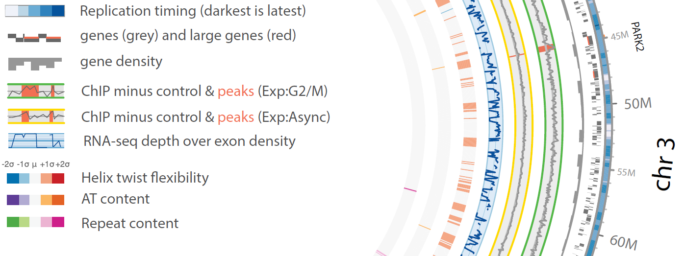
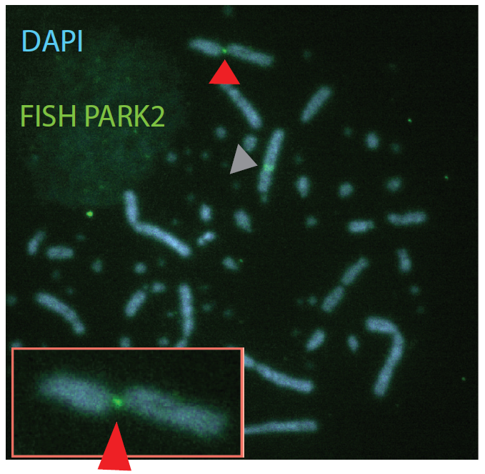
Chromosomal fragile sites (CFSs) are genomic regions prone to form gaps or breaks on metaphase chromosomes. As CFSs often appear as hot spots for gross chromosomal rearrangements in cancer cells, it is of high clinical relevance to map CFSs genome-wide for better understanding of complex aberrant karyotypes and to predict their clinical prognosis. For now, CFSs have mainly been analysed in human lymphocytes but the method is also established for other tissues and model systems. The mapping usually followed the traditional method by metaphase spreading and molecular cytogenetic definition. A new approach has become available recently to standardize this mapping to a high throughput and high resolution Next Generation Sequencing (NGS)-based genome-wide application. The aim of my junior research project is to establish this novel method in human cells, implement it for tumor material to overcome conventional metaphase spreading and morphological definition by a state-of-the-art NGS approach. To expand the view on CFSs by specific functional analyses in disease-derived cell lines, aspects of nuclear localization as well as molecular positioning of CFSs will be investigated. Overall, the project aims to identify the connection between the fragility of certain genomic regions and cancer predisposing and premature aging related disorders.

<b>Figure 1:</b> Genome-wide FANCD2 binding analysis, partially depicted circos plot of Gallus gallus chromosome 3 indicating replication timing, gene position, gene density, FANCD2 ChIP-seq results, transcription levels and DNA sequence features. One of the most significant peaks is residing in the PARK2 gene. (from Pentzold et al., 2017)
Figure 1: Genome-wide FANCD2 binding analysis, partially depicted circos plot of Gallus gallus chromosome 3 indicating replication timing, gene position, gene density, FANCD2 ChIP-seq results, transcription levels and DNA sequence features. One of the most significant peaks is residing in the PARK2 gene. (from Pentzold et al., 2017)
 
<b>Figure 2:</b> Metaphase spread from chicken DT40 cells, treated with the replication inhibitor aphidicolin. The FISH probe hybridizes to PARK2, which is broken (close up image in red box and red arrowheads). One allele of PARK2 is not broken (grey arrowhead). (from Pentzold et al., 2017)
Figure 2: Metaphase spread from chicken DT40 cells, treated with the replication inhibitor aphidicolin. The FISH probe hybridizes to PARK2, which is broken (close up image in red box and red arrowheads). One allele of PARK2 is not broken (grey arrowhead). (from Pentzold et al., 2017)
 
Dr. Constanze Pentzold
Molekulargenetische Diagnostik,
Arbeitsgruppenleiterin

Telefon: +49 3641 9-396861
Web
Homepage Genetical counseling Publications
General Information Diagnostic accompanying letters Model Project Genome Seq
Fields of work Teaching
Clinical diagnostics Databases
Europäischer Sozialfonds
Europäische Union - Sozialfonds
TMWWDG
EFRE-EU
Akkreditierungsurkunde Instititut für Humangenetik
Akkreditierungsurkunde Praxis für Humangenetik - ZAM
FSU
VUD