Zur Startseite Zur Hauptnavigation Zum Inhalt Zum Kontakt Zur Suche Zur Sitemap
+A-
ENDE
  menu
Summer School
  menu
  • Home
  • Program 2025
  • Program 2024
    • Module Complex I
      • Module I/1
      • Module I/2
      • Module I/3
      • Module I/4
      • Module I/5
      • Module I/6
    • Module Complex II
    • Module Complex III
    • Module Complex IV
  • Application
  • Jena
  • Contact
  • Links
  • News
Summer School Molecular Medicine / Program 2024 / Module Complex I

Module I/1 - Novel technologies in leukemia research

Prof. Dr. Thomas Ernst
Department of Internal Medicine II,
Haematology/Oncology
Am Klinikum 1
07747 Jena

phone: +49 3641 9-327119
Web

Chronic myeloid leukemia (CML) represents a model disease for cancer biology, diagnostics and
treatment. CML became well treatable with the approval of tyrosine kinase inhibitors (TKIs)
which selectively block the oncogenic fusion protein BCR-ABL1. Many patients achieve deep
molecular remission and may even stop therapy.
A wide range of leukemia associated genes are investigated routinely in our lab by next-generation
sequencing. Single cells obtained from leukemia patients are analyzed to investigate the interaction
and sequential acquisition of mutations as a rationale for novel treatment strategies. With the highly
sensitive droplet digital PCR, which distributes the sample over 20,000 individual nanoliter-sized
droplets, our group attempts to identify those CML patients, who can safely discontinue therapy.
During the course, we will provide hands-on training from blood preparation to high-throughput
genotyping using novel sequencing technologies (e.g. next-generation sequencing, pyrosequencing).
Students will learn how to pick single cells, perform mutation-specific PCRs, DNA sequencing and
droplet digital PCR. In addition to these experimental techniques students will learn different types
of bioinformatic tools.

Fig 1 (left) Detection of BCR-ABL fursion transcription by digital PCR.     
Fig. 2 (right) Identification of a homozygous Jak2 V617F point mutation in HEL cell.  

Module I/2 - Investigations on diabetic nephropathy

Dr. Tzvetanka Bondeva
Department of Internal Medicine III ,
Nephrology Lab
Am Klinikum 1
07747 Jena

phone: +49 3641 9-324354
Web

The MAPK-organizer protein 1 (MORG1), plays a scaffolding function for number of molecules involved in the MAPK cascade. Furthermore, it was found that it binds to prolyl-hydroxylase -3 (PHD3) isoform. Our studies revealed that reduction of MORG1 expression leads to reduced PHD3 activity and increased HIFs transcriptional activation. While MORG1 KO mice are embryonic lethal, the reduction of MORG1 expression in MORG1+/- mice was associated with an increased stabilization of the HIFs in animal models of Type 1 and 2 DM, as well as in ischemia-reperfusion model, compared with equally treated wild type MORG1 animals. This resulted in a decreased apoptosis, reduced inflammation and improved renal function. Furthermore, we observed that a reduced expression of MORG1 has a renoprotective effect in animal models of short-time systemic hypoxia and endotoxemia. Therefore, understanding the molecular mechanisms of the transcriptional regulation of MORG1 expression and the protein complexes in which MORG1 is involved in renal cells could be of therapeutic interest.  

During the summer school experimental week we will study the effects of different factors, as increased glucose levels, TGF-beta, hypoxia or 3,4 DHB (PHDs inhibitor) on the MORG1 expression in HEK 293 cells. Using as a tool CRISPR/CAS9 system, we generated HEK 293 cells, expressing human MORG1-fusion to Luciferase gene in order to monitor the Luciferase activity as a reporter for the MORG1-protein expression. Furthermore, the MORG1 expression will be assayed by real – time PCR. The cellular distribution of MORG1-protein will be visualized by immunofluorescent staining in HEK293 cells and/ or murine renal cells.

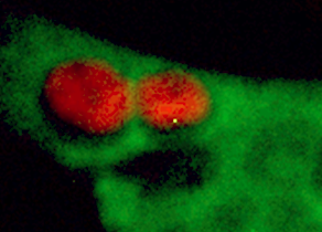
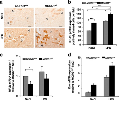
Figure 1 a-d: Reduced expression of MORG1 in MORG1+/- heterozygous mice is associated with an elevated renal expression of HIF-2a protein, respectively an increased mRNA levels of the HIF-2a target gene erythropoietin (Epo)  in LPS treated mice, compared to the MORG1+/+ wild type endotoxemic mice.

Bondeva T., Schindler C., Schindler K., Wolf, G. MORG1(+/-) mice are protected from histological renal damage and inflammation in a murine model of endotoxemia. BMC Nephrol. 2018 Feb 5; 19 (1):29.


Module I/3 - Analysis of hormone-induced cellular senescence

Prof. Dr. Aria Baniahmad
Institute of Human Genetics
Am Klinikum 1
07747 Jena

Web

Cellular senescence leads to an irreversible block of cellular division capacity (G0-Phase of cell cycle) both in cell culture and in vivo. The induction of an irreversible cell cycle arrest is very useful in the treatment of cancer. Therefore, targeting cellular senescence may represent a new approach for cancer therapy. However, senescent cells exhibit the senescence-associated secretory phenotype (SASP) that may influence other neighboring cells and cancer cells in their differentiation and proliferation.

Cellular senescence is associated with a dramatic change in nuclear architecture observed by formation of heterochromatic foci. It is also associated with the expression of cell cycle inhibitors such as p15, p16, ARF and p21. The detailed molecular mechanisms of induction of cellular senescence are unknown.

Interestingly hormones may induce cellular senescence in cancer cells. Specifically, we will use human prostate cancer cells. Prostate cancer is a major age-related disease and the second cause of cancer death in males. Androgen-dependent and castration resistant prostate cancer cells will be used. Importantly, both cell types are regulated by the androgen receptor signaling. Both cell types will be treated with androgens or androgen-receptor antagonists. Senescence specific markers will be analyzed including chromatin changes. Further, Western blotting and real-time PCR will be employed for detection of changes of key factors involved in cellular senescence.


Module I/4 - Studying gene regulation with modern techniques

Prof. Dr. Ralf Mrowka
Department of Internal Medicine III,
Experimental Nephrology
Am Nonnenplan 4
D-07743 Jena

phone: +49 3641 9-396600
fax: +49 3641 9-396602
Web

The Group Experimental Nephrology is focused on investigating gene regulation in the context of the kidney, but also of transcription factors and immunological genes. We functionalize human cell lines with modern genetic techniques like CRISPR/Cas9, FlpIn/FRT and TALEN to use them in luciferase or fluorescence reporter assays.

We study receptor activation in innate immune system activation. Those pattern recognition receptors recognize a wide range of molecular patterns. Their activation recruits and activates signal cascades leading to the translocation of transcription factors from plasma to the nucleus. The transcription of inflammation-specific genes is activated and induces the innate immune response.

The generation of cell-based toxicity reporter systems in human cell lines and induced pluripotent stem cells (iPS cells) is a possibility to study organ-specific toxicity reactions. We differentiate iPS cells into different cell types and monitor changes in the cell identity by reporter gene activation and analyze cell function by FACS, fluorescence microscopy and molecular biology techniques.

keywords: genetic modification, cloning, transient and stable transfection, fluorescent microscopy, luminometry

Figure: Left: Life cell imaging of a transcription factor (green) and a nuclear protein (red) right: Simplified principle of reporter gene activation after stimulation of a TLR pathway

Module I/5 - Developing mass spectrometry technologies to decipher the proteome of cancer cells

Prof. Dr. Florian Meier (Jun.-Prof.)
Jena University Hospital,
Functional Proteomics
Am Klinikum 1
07747 Jena

phone: +49 3641 9-390879

The era of personalized medicine promises data-driven therapeutic strategies that are tailored to individual patients based on molecular profiles. From a functional perspective, proteins are the key drivers of our cells, which also makes them primary targets of molecular therapies. Collectively, they build the proteome, which orchestrates virtually any biochemical process from energy conversion to signal transduction and the cell cycle. Conversely, aberrant protein homeostasis and signaling are hallmarks of many diseases, including cancers.

The goal of proteomics is to study the proteome in its entirety and in a quantitative, unbiased manner. Mass spectrometry (MS)-based proteomics has evolved as the method of choice for many applications. It has proven successful in the stratification of cancer subtypes from tissue biopsies as well as in disentangling cascades of post-translational modifications in signaling networks. This has been enabled by tremendous advances in the proteomics technology, which remains a driving force as the field strives towards analyzing large cohorts of clinical sample and single cells.  

Our research focuses on new techniques in mass spectrometry to increase throughput, sensitivity and selectivity of the analysis. We employ latest-generation trapped ion mobility – time-of-flight mass spectrometry to analyze proteins and post-translational modifications of patient-derived cells, biofluids and tissue samples.

The summer school will introduce you to the fundamentals of MS-based proteomics. The course covers biochemical techniques to prepare samples from human specimens and process them for mass spectrometric analysis. We will make use of the latest technology to acquire quantitative data for several thousand proteins in a single experiment. Students will gain insight in different acquisition strategies and multi-dimensional separation techniques, including liquid chromatography and ion mobility spectrometry. We will use bioinformatics tools to analyze raw data and interpret the results.

Module 1/6 - Introduction into the analysis of omics data using R

Prof. Dr. Rainer König
Institute for Infectious Diseases and Infection Control,
Systemsbiology Research Group
Kollegiengasse 10
07743 Jena

phone: +49 3641 9-397830
Web

Most often, the disease or cellular condition under study has been investigated in some related context by others having performed transcritional profiling or other sequencing based investigations (genomics, ChIP-seq, epigenomics), Crispr/Cas9 or RNAI knockout/down screens or protein mass spectrometry. According to the FAIR principles (Findable, Accessible, Interoperable, Reusable), such data should be accessible, and, indeed this is often the case. Interrogating such data may be a useful shortcut to bring up or confirm own hypotheses. However, having a bioinformatician at hand may turn out to be the bottleneck. Hence, learning the basics for analysing such omics data can be useful also for experimentally working biomedical scientists!
We will introduce you into the R programming language. You will learn how to write small computer programs enabling to download such omics data and apply basic analysis and statistical methods. Exemplarily, we will download transcriptomics data, e.g. from an infectious disease, such as from COVID19 patients or a disease caused by another pathogen. The data will be normalized, differentially expressed genes identified, genes set enrichment tests performed to elucidate pathways being affected by the disease, clustering to find similarly regulated genes or samples (from e.g. patients) and classification performed to construct a diagnosis tool.
You do not need any prior knowledge in programming and will learn how to use such an analysis pipeline from scratch.

Home Jena
Program 2025 Contact
Program 2024 Links
Application News
Summer School
DAAD
IZKF