Zur Startseite Zur Hauptnavigation Zum Inhalt Zum Kontakt Zur Suche Zur Sitemap
+A-
ENDE
  menu
Summer School
  menu
  • Home
  • Program 2025
  • Program 2024
    • Module Complex I
    • Module Complex II
    • Module Complex III
      • Module III/1
      • Module III/2
      • Module III/3
      • Module III/4
      • Module III/5
      • Module III/6
    • Module Complex IV
  • Application
  • Jena
  • Contact
  • Links
  • News
Summer School Molecular Medicine / Program 2024 / Module Complex III

Module III/1 - Live cell imaging - watching nerve cells in action

Prof. Dr. Britta Qualmann
Institute of Biochemistry I
Nonnenplan 2
07743 Jena

phone: +49 3641 9-396300
Web
PD Dr. Michael Kessels
Institute of Biochemistry I
Nonnenplan 2
07743 Jena

phone: +49 3641 9-396310
Web

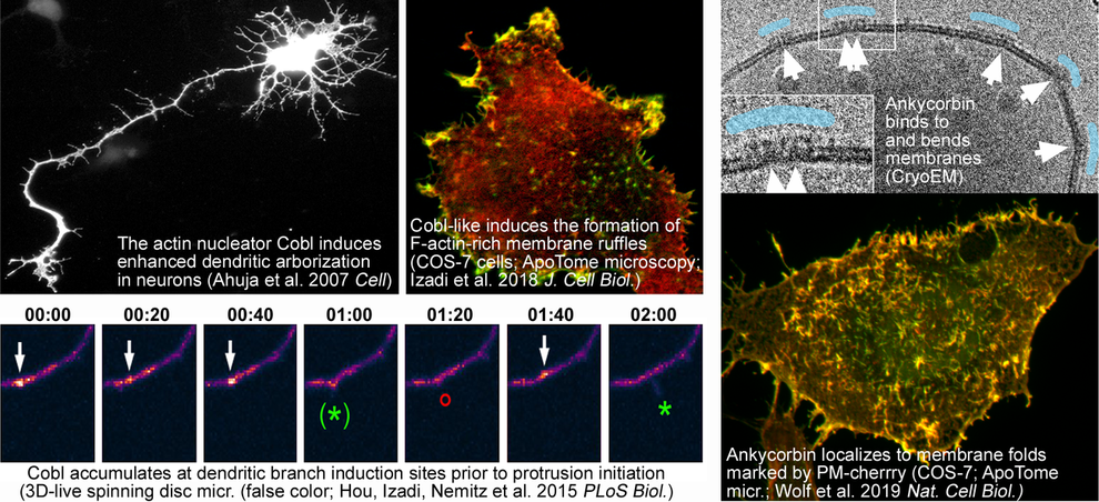
A prerequisite for establishment of cellular morphology is the ability of cells to remodel their plasma membrane and to reorganize the actin cytoskeleton in response to external and internal cues. In neurons, actin dynamics and membrane remodelling are indispensable for neuronal network formation, for information processing in the central nervous system and for learning and memory processes. Alterations in dendrite morphology or defects in neuronal development contribute to several neurological and neurodevelopmental disorders, such as autism spectrum disorders, Alzheimer's disease, schizophrenia, Down syndrome and depression.

A major research focus of our groups therefore is to understand how reshaping of the plasma membrane is elicited by proteins, which directly interact with and/or deform the lipid bilayer (membrane shapers), and this is promoted by dynamics of the cortical actin cytoskeleton.

In the summer school, we will apply modern techniques in molecular biology, cell biology and microscopy to study the organization and dynamics of the actin cytoskeleton, effector molecules and Ca2+-signals. Imaging neuronal cell dynamics reveals how structural and functional cellular plasticity is brought about. These processes are indispensable for adaptations of neuronal contacts and reorganizations underlying brain development, regeneration, learning and memory.

Modern molecular tools will allow for a state-of-the-art analysis of cytoskeletal structures at high resolution. Students will have the opportunity to gain hands-on expertise in live cell imaging using apotome-based confocal microscopy as well as spinning disk microscopy. High-resolution 3D-live analyses will be used to track cellular morphology and proteins implicated in neuronal network formation and nerve cell communication in time and space and to study their dynamics.

The studies in the course of this summer school project will thereby shed light on the fascinating cellular adaptations underlying development, plasticity and information processing/learning in vertebrate brains and on how these functions are derailed in pathological and disease states.

Module III/2 - Synaptic plasticity in neurological disease

Prof. Dr. Christian Geis
Hans Berger Department of Neurology,
Section of Translational Neuroimmunology
Am Klinikum 1
07747 Jena

phone: +49 3641 9-323413
Web

The research interests of our group focusses on autoimmune mechanisms on central synaptic pathology.

Recent findings in patients with autoimmune encephalitis showed that these disorders are associated and presumably induced by highly specific autoantibodies against synaptic target structures, e.g. ionotropic receptors (NMDA, AMPA, Glycin receptors), G-protein coupled receptors (e.g. GABA-B receptors), or synaptic linker proteins (e.g. LGI1). The group of C. Geis investigates the effects of human autoantibodies on pre- and postsynaptic mechanisms. We have established passive-transfer mouse models with application of purified patient antibodies that are used to study the pathogenic effects of antibodies on animal behavior. Moreover, autoantibody-induced synaptic dysfunctions are evaluated in neuronal cell cultures, acute brain slices, and in-vivo using neurophysiological techniques. Super-resolution imaging techniques are used to investigate distinct antibody-induced morphological changes on the nanoscale level.

Another research focus is on pathomechanisms involved in sepsis associated encephalopathy. Here, the group of C. Geis uses a murine peritoneal contamination and infection model (PCI) to evaluate immune-mediated processes that may cause neuronal damage underlying cognitive deficits. Of particular interest is the role of microglia as the brain’s resident immune cells. Techniques used involve qPCR and RNASeq to analyze changes on the transcriptome level as well as immunohistochemical stainings of brain slices for morphological analyses. Several behavioural tests are performed to evaluate neurocognitive deficits after PCI. 

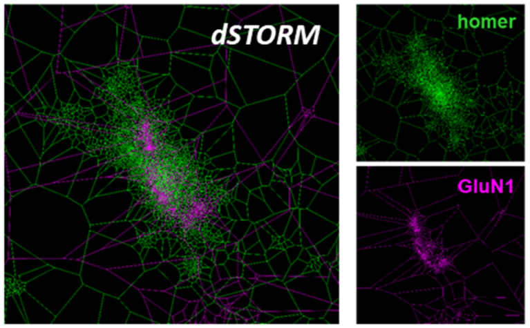
„Binding of human antibodies from a patient with NMDA receptor encephalitis to the GluN1 subunit
of the NMDA receptors as shown by super-resolution dSTORM imaging and tesselation cluster
analysis
Upper right picture: Homer staining; homer protein clusters and colocalize with NMDA receptors
right picture below: Same synapse, GluN1 staining
Left picture: overlay of homer and GluN1 staining

Module III/3 - Imaging of morphological and functional plasticity in the rodent brain

Dr. Anja Urbach
Hans Berger Department of Neurology
Am Klinikum 1
07747 Jena

phone: +49 3641 9-32 59 01
Dr. Silvio Schmidt
Hans Berger Department of Neurology
Am Klinikum 1
07747 Jena

Learning and adaptation are fundamental properties of the brain that are based on the continuous remodelling of neuronal networks. Our research group at the Department of Neurology investigates cellular and molecular mechanisms underlying dynamic structural and functional changes in the young, aging and diseased brain, focusing on adult neural stem cells and neurogenesis, as well as neuronal and glial plasticity. Therefore, we employ modern neuroimaging methods (e.g. magnetic resonance imaging, confocal laser microscopy), state-of-the art molecular techniques (e.g. single-cell sequencing, RiboTag), bioinformatics and behavioural testing.  

In the upcoming summer course, we will introduce our research topics as well as (i) interventions to induce plasticity, (ii) behavioral tests to assess functional outcomes, (iii) imaging techniques to visualize plasticity in the brain and (iv) molecular biological and histological methods to investigate cellular changes in plastic brain areas. In addition, you will gain practical experience with cell type-specific RNA extraction, quantitative PCR, immunohistochemical methods and confocal laser microscopy.


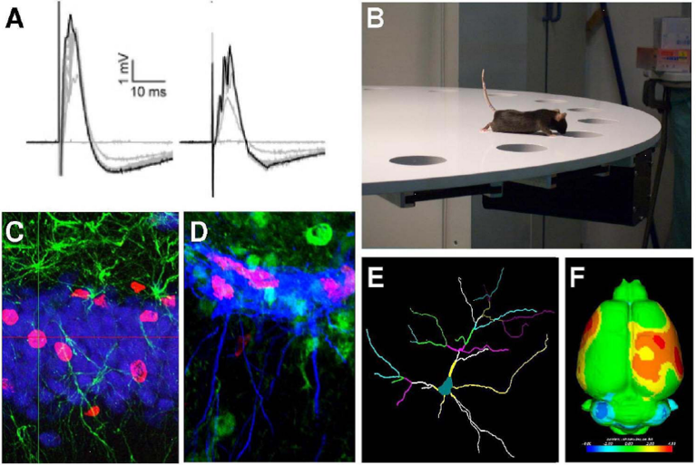
A) Electrophysiological recordings of stimulus-dependent responses in 2 different areas of the hippocampus. B) Mouse passing the Barnes maze test. C) Newborn neurons (purple) and D) newborn neuronal precorsor cells (purple) in the adult dentate granule layer. E) Tracing of a neuron residing layer II-III of the mouse motor cortex. F) MRI image of volumetric changes in rat model of cortical malformations.

Module III/4 - Introduction to fluorescence lifetime imaging (FLIM)

Prof. Dr. Christoph Biskup
Biomolecular Photonics Group
Nonnenplan 4
D-07743 Jena

Web

In conventional fluorescence microscopy only the information conferred by the fluorescence intensity is exploited. But also other intrinsic properties of a fluorophore such as the fluorescence lifetime can be used to retrieve additional information about the sample. Fluorescence lifetime imaging (FLIM) can report on photophysical events that are too difficult (or impossible) to observe by fluorescence intensity measurements.

Since the lifetime of a fluorophore depends on its local environment it can be used as a sensor for properties of the solvent such as the viscosity and dissolved compounds such as ions or oxygen. By exploiting physical phenomena such as Förster energy transfer (FRET), FLIM can be used to test if molecules are in close vicinity to each other. In this way molecular interactions can be “imaged”.

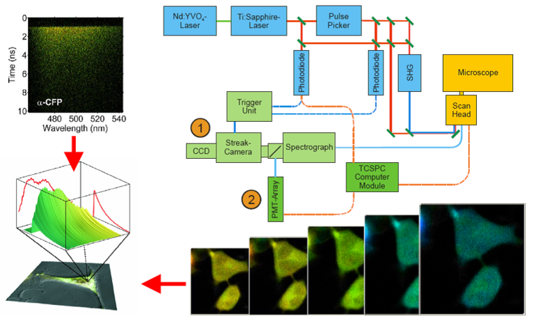
In the lab of the Biomolecular Photonics Group several methods to measure fluo­res­cence lifetimes are established. One tech­nique is based on time-correlated single photon counting (TCSPC); the other method is based on a streak-camera system. This course will give an introduction into the technical equip­ment necessary to acquire the data and the mathe­matical algorithms used to analyze the data.

Further information about the course can be found on the website of the Biomolecular Photonics group: https://www.uniklinikum-jena.de/photonik/en/Teaching/Summer+School.html   


Setup used for fluorescence lifetime mea­sure­ments. A pulsed Titanium:Sapphire (TiSa) laser or a super­continuum laser are used for pulsed excitation of the biological sample on the stage of a confocal laser scanning microscope. Fluorescence emitted by the sample is recorded with a streak-camera (1) or a fast PMT-array (2) with a high spatial (x,y), temporal (t) and spectral (l) resolution.

Module III/5 - In vivo small animal imaging of disease-related molecular structures

Prof. Dr. Ingrid Hilger
Institute of Diagnostic and Interventional Radiology,
Experimental Radiology Group
Am Klinikum 1
07747 Jena

phone: +49 3641 9-325921
fax: +49 3641 9-325922
Web

This course deals with the potential and the feasibilities of in vivo imaging of disease-related molecular markers in small laboratory animals. Namely, in vivo molecular imaging has been established to be a critical component of preclinical and translational biomedical research. It allows researchers to determine the biological structure and function of molecular markers by non-invasive means in situ in the body, i.e. without the withdrawal of tissue from the body for further analysis. This means that quantitative, spatial and temporal information on normal and diseased tissues can be determined, for example of those associated with cancer, inflammation or neurodegenerative diseases. Particular aims of such research activities are the discovery and analysis of disease-associated molecular interrelations, the elucidation of dedicated therapeutic effects, and validation of new drugs in the in vivo situation. There are several imaging modalities available for small animal imaging (e.g. mice and rat), such as whole body near infrared optical imaging, computed tomography (CT), positron emission tomography (PET), single photon emission tomography (SPECT), magnetic resonance tomography (MR), etc.

Figure 1. Enrichment of three different nanoparticle formulations into tumors. Representative composite macroscopic near infrared fluorescence and light pictures of the back of mice bearing CAL-27 tumors. From Haedicke et al. 2015, Acta Biomaterialia 14, 197.

In this course students will have the opportunity to get insights into: 1) the principles of the most important small animal molecular imaging modalities, 2) the construction of molecular imaging probes, 3) the basic requirements for animal experimentation, 4) the imaging of a local inflammation by utilization of small animal molecular imaging modalities, 4) the analysis of probe pharmacokinetics and the definition of physiological barriers counteracting to probe accessibility, and finally 5) the identification of potential pitfalls in image interpretation. Methods will cover areas concerning fluorescence microscopy, cytology, cell culture, optical spectroscopy, protein chemistry, pharmacology, disease models in mice, macroscopic optical imaging, CT imaging, histology, data analysis and statistics, etc.

Hands-on experience will be guided by experienced staff of the “Experimental Radiology Group”. All demonstrations and experimentation will be performed on state of the art devices and complemented with lectures on molecular imaging technology.

Module III/6 - Visualization of GPCR signal-transduction in real-time

Prof. Dr. Carsten Hoffmann
Institute of Molecular Cell Biology,
Center for Molecular Biomedicine
Hans-Knöll-Str. 2
07745 Jena

phone: +49 3641 9-395600

In order to transduce a signal of a hormone or prescription drug across the plasma membrane G-protein-coupled receptors (GPCRs) need to undergo conformational changes. The focus of our research is to investigate such conformational changes during GPCR activation and deactivation. Therefore we develop FRET-based probes for GPCRs to image the conformational change in living cells and millisecond time resolution. The use of such FRET-based sensors allows us to study receptor ligand interaction directly at the level of the receptor itself. Thus we are able monitor the effects of potential future drugs at the protein level and can correlate the observed data with effects on different signalling pathways triggered by receptor activation.

Receptor interaction with b-arrestin is an important regulatory key element in the termination of G-protein-dependent receptor signalling. The interaction of a GPCR and b-arrestin is regulated by ligand binding and receptor phosphorylation by specific receptor kinases. Since β-arrestins not only turn off G-protein-dependent-signalling but represent starting points for novel signalling cascades, we are also interested to investigate receptor ligands which are able to discriminate between G-protein and b-arrestin mediated receptor signalling. Such compounds are called biased ligands and are of great value for basic research and hold the promise for fewer side effects for patient treatment.

During the Summer School, we focus on real-time analysis of receptor activation in living cells using FRET based bio-sensors for the signal transduction pathways involved in receptor signalling. Applied techniques will include heterologous expression in mammalian cells, plate-reader assays and microscopy of signal transduction in living cells.

Home Jena
Program 2025 Contact
Program 2024 Links
Application News
Summer School
DAAD
IZKF