Zur Startseite Zur Hauptnavigation Zum Inhalt Zum Kontakt Zur Suche Zur Sitemap
+A-
ENDE
  menu
Summer School
  menu
  • Home
  • Program 2025
    • Module Complex I
    • Module Complex II
      • Module II/1
      • Module II/2
      • Module II/3
      • Module II/4
      • Module II/5
      • Module II/6
      • Module II/7
    • Module Complex III
    • Module Complex IV
  • Program 2024
  • Application
  • Jena
  • Contact
  • Links
  • News
Summer School Molecular Medicine / Program 2025 / Module Complex II

Module II/1 - Effects of preconditioning on cardiomyocytes and embryonic stem cells

Prof. Dr. P. Christian Schulze
Department of Internal Medicine I
Am Klinikum 1
07747 Jena

Web
Dr. Mohamed M. Bekhite ELsaied
Department of Internal Medicine I
Am Klinikum 1
07747 Jena

Web

Topic 1 - Analysis of cardiac lipid metabolism (Prof. Dr. P. Christian Schulze)

The heart derives most of its ATP generating substrates from fatty acids (70%) and glucose with small amounts of ketons bodies, lactate and amino acids under physiologic conditions. Under cardiac stress such as ischemia and hypertrophy or failure, cardiac metabolism switches to glycolytic utilization of glucose as the primary source for ATP generation. This creates a metabolic deficit and energy depletion of the failing heart. Further, normal oxidative metabolism and mitochondrial function is impaired leading to accumulation of toxic lipid intermediates such as ceramides and other sphingolipids. The current project studies the contribution of various sources of metabolites to cardiac metabolism in the normal and failing heart and in isolated cardiomyocytes and fibroblasts. We use transgenic models of ceramide synthase overexpression and deletion tos study metabolic flux in vivo and in vitro.

Topic 2 - Induced Pluripotent Stem (iPS) cells as in vitro model of human heart

The discovery of induced pluripotent stem (iPS) cell technology has opened up unprecedented opportunities to generate patient-specific cell types in vitro, to elucidate the underlying mechanisms of physiology and pathology of the human heart. For that, we established the human-iPS cell technology as a promising generation of pluripotent stem cells using non-integrating methods to introduce the reprogramming factors (OCT4, SOX2, KLF4, and c-MYC) to the somatic cells.

The generation of human-iPS cell-derived cardiomyocytes is of growing interest for multiple applications. First, access to an in vitro model of human development permits the study of human heart development. Second, iPS cell-derived cardiomyocytes serve as a human cardiac model that can be used for diverse basic research studies ranging from cellular electrophysiology to protein biochemistry. Furthermore, human-iPS cells represent a potential source of cell-based therapies to repair and replacement of diseased or damaged tissues.

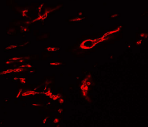
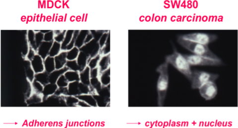
The purpose of this course to learn the basics of human-iPS cell reprogramming, iPS cell culture and differentiation of human-iPS cells into cardiovascular tissue. Therefore, we will analyze the changes in cellular and molecular of normal human-iPS cells-derived cardiovascular tissue compared with diseased tissue using different methods, such as calcium transient quantification, Seahorse metabolic profiling, gene expression, protein level, flow cytometry, morphological analysis of mitochondria, immunofluorescence staining and examined using a confocal laser scanning microscope (cLSM 900).

Module II/2 - Signal transduction in leukemic cells

Prof. Dr. Jörg Müller
Institute of Molecular Cell Biology,
Center for Molecular Biomedicine
Hans-Knöll-Str. 2
07745 Jena

phone: +49 3641 9-395634

Acute Myeloid Leukemia comprises a heterogeneous group of severe diseases with limited treatment possibilities, especially in elderly patients. Therefore, intense research aims at identifying novel drug targets and related therapies. Mutations giving rise to the oncoprotein FLT3 ITD (Fms-like tyrosine kinase with internal tandem duplications) represent one of the important classes of driver mutations in a subset of 25 – 30 % of patients. FLT3 is a member of the class III receptor tyrosine kinases and is involved in cell survival, proliferation, and differentiation of haematopoietic progenitors of lymphoid and myeloid lineages. Targeting FLT3 mutant leukemic stem cells (LSC) is a key to efficient treatment of patients with relapsed/refractory AML.
We will carry out experiments targeting the oncogenic activity of mutant FLT3 proteins.

Figure: FLT3-mediated Signaling

Müller JP, Schmidt-Arras D. Novel Approaches to Target Mutant FLT3 Leukaemia. Cancers (Basel). 2020 Sep 29;12(10):2806.

Module II/3 - Signaling molecules in angiogenesis

Prof. Dr. Regine Heller
Institute of Molecular Cell Biology,
Center for Molecular Biomedicine (CMB),
Jena University Hospital
Hans-Knöll-Str. 2
07745 Jena

phone: +49 3641 9395633
The figure shows blood vessel formation in Matrigel plugs in mice in response to basic fibroblast growth factor. Matrigel plugs were fixed in Zinc fixative and embedded in paraffin. 5 µm sections were prepared and stained for the endothelial marker protein CD31.

Angiogenesis, the growth of blood vessels, is an essential biological process under both physiologic and pathologic conditions. It requires the tight coordination of endothelial cell proliferation, differentiation, migration, of matrix degradation and of cell-cell signaling processes. Angiogenesis is controlled by a variety of growth factors with Vascular Endothelial Growth Factor (VEGF) being one of the most important molecules.

Our interest focuses on the characterization of signalling pathways in endothelial cells in response to proangiogenic stimuli, in particular to VEGF. We are especially interested to understand how signals initiated or controlled by oxidative or metabolic stress affect endothelial cell functions. In this context, we investigate the role of the energy sensor AMP-activated kinase (AMPK) and of oxidant-sensitive signaling molecules such as protein tyrosine phosphatases. Recently, we found that AMPKα1 plays an essential role in VEGF-induced angiogenesis in vitro and in vivo and characterized the VEGF/AMPK/GFAT* pathway as one of the responsible mechanisms  (* GFAT - glutamine fructose-6-phosphate amidotransferase; Stahmann N et al. J Biol Chem. 285:10638-52, 2010, Zibrova D et al. Biochem J 474:983-1001, 2017).

The course will provide hands-on training in preparing primary endothelial cells of human or mice origin and in performing different migration and angiogenesis assays (transwell migration, wound healing assays, spheroid assays, matrigel assays). These tests involve cellular fluorescence microscopy and the ECIS (Electric Cell-Substrate Impedance Sensing) technology. Further, the students will get familiar with RNA interference techniques and shRNA lentiviral transduction in primary cells to investigate the role of selected molecules in angiogenesis.

Module II/4 - Studying cooperativity in ionotropic and metabotropic receptors

Prof. Dr. Klaus Benndorf
Institute of Physiology II
Kollegiengasse 9
07743 Jena

phone: +49 3641 9-397651
Web
Dr. Ralf Schmauder
Institute of Physiology II

Web

Cells need to adapt and fine-tune to varying environmental stimuli. Receptors in the plasma membrane translate chemical stimuli to changes in membrane potential or activation of signaling cascades. This module will offer an insight into different strategies to study the initial states of this process: the ligand binding an activation of the receptor. Often, receptors are multi-subunit proteins. This allows the emergence of cooperativity and thus a fine tuned sensitivity, which we will study here.

In the case of ligand-gated ion channels, also called ionotropic receptors, the controlling stimulus is the binding of ligand molecules to intracellular or extracellular binding sites of the channel protein.  A standard approach to characterize their activation behavior in response to a ligand stimulus is the electrophysiological patch-clamp technique. Additionally, you will learn strategies to use fluorescently labeled ligands to follow receptor affinity and cooperativity, with fluorescence microscopy and confocal patch-clamp fluorometry (confocal PCF). In this method, we combine patch clamp and confocal microscopy to simultaneously study ligand binding and channel activation.

For metabotropic receptors, the activation is monitored by following conformational changes with FRET between to fluorophores engineered into the receptor.

Finally, you will learn strategies to generate quantitative models from the obtained data and draw conclusion on receptor cooperativity. 


A) Scheme of a patch-clamp setup. The illustration shows the inside-out configuration with a glass pipette containing a membran patch excised from a Xenopus laevis oocyte.

B) Confocal image of a patch-pipette with an excised membrane patch expressing a high density of CNGA2 channels. The green fluorescence signal is due to the binding of fluroescently labelled cGMP molecules to the ion channels’ binding sites.

Module II/5 - ß-Catenin signaling in disease

Prof. Dr. Otmar Huber
Institute of Biochemistry II
Nonnenplan 2
07743 Jena

phone: +49 3641 9-396400
Web

ß-Catenin originally was identified as a component of the cadherin-catenin cell adhesion complex. Genetic experiments revealed a second function of ß-catenin within the Wnt signaling pathway. In unstimulated cells free ß-catenin is rapidly degraded by the ubiquitin-proteasome pathway.

Binding of secreted Wnt proteins to Frizzled-LRP5/6 receptor complexes activates the canonical Wnt pathway. This results in the inhibition of the ß-catenin destruction complex and an intracellular accumulation of ß-catenin. Within the nucleus ß-catenin regulates transcription of target genes in binding to LEF/TCF transcription factors. Deregulated activation of this pathway by mutations of pathway components affects cell proliferation, differentiation and apoptosis. We are interested in the molecular mechanisms that modulate ß-catenin function. During recent years we identified new ß-catenin interaction partners using co-immunoprecipitation and pull-down assays with purified recombinant proteins and mapped the corresponding binding sites. To study the functional relevance of these new interaction partners, we use a broad spectrum of methods including reporter gene assays, real-time RT-PCR, ChIP and two-step ChIP, shRNA, cell proliferation, cell migration and anchorage independent growth experiments.


In MDCK cells ß-catenin is localized at adherens junctions in association with E-cadherin. In SW480 colon carcinoma cells, a mutation in the APC tumor suppressor protein, which is a component of the ß-catenin destruction complex, results in cytoplasmic and nuclear accumulation of ß-catenin.

Module II/6 - Unlocking the potential of retinoic acid in anticancer therapy

Dr. Tino Schenk
Department of Internal Medicine II,,
Haematology/Oncology
Hans-Knöll-Str. 2
07745 Jena

phone: +49 3641 9-395637
Web

Acute myeloid leukemia (AML) encompasses a cytogenetically heterogeneous group of myeloid malignancies characterized by clonal expansion of abnormally or poorly differentiated myeloid cells in the bone marrow, blood and other tissues. Treatment failure, relapse and mortality of AML is unacceptably high and it is reasonable to predict that cure rates will not improve unless treatment modalities alternative to conventional chemotherapy and bone marrow transplantation are developed. A hallmark of AML is a failure to properly differentiate and the retinoic acid receptor (RAR) ligand all-trans-retinoic acid (ATRA) has demonstrated remarkable efficacy in inducing differentiation in a sub-type of AML, acute promyelocytic leukemia (APL). However, ATRA based treatment has failed to replicate this success in non-APL AML.

Our research is aimed towards the identification and characterization of mechanisms underlying the maturation arrest and ATRA-resistance of leukemic cells eventually enabling efficient therapy by induction of differentiation in all types of AML. We especially focus on epigenetic factors preventing the induction of myeloid differentiation by ATRA. Currently we investigate the roles of the lysine demethylase LSD1 and the lysine acetyltransferase GCN5.

Within this course we will test the effects of compounds inhibiting epigenetic modifiers as LSD1 and GCN5 on AML cells. We will assess cell growth using cell viability assays and perform FACS analyses measuring changes in myeloid differentiation and apoptosis markers following treatments. Further, we will perform basic analyses of RNA-seq data previously obtained.


Module II/7 - Foundation of life: cellular and molecular communication in the placenta

PD Dr. Diana Maria Morales Prieto
Department of Obstetrics,
Placenta Lab
Am Klinikum 1
07747 Jena

phone: +49 3641 9-390859
Web
Dr. José Martin Murrieta Coxca
Department of Obstetrics,
Placenta Lab
Am Klinikum 1
07747 Jena

phone: +49 3641 9-329292
Web

From an immune point of view, the pregnancy represents a complex process involving intricate interactions between the mother's immune system and the developing fetus. The assumption that entities foreign to a specific organism should be rejected by its immune system is challenged by the fact that despite expressing foreign paternal antigens, the fetus remains in the mother’s uterus instead of being rejected in the way that graft would be. The placenta appears as the interface between the mother and the fetus and is the organ that modulates their communication in a cellular and molecular way. The placenta creates a barrier that regulates the contact between maternal and fetal cells, preventing immune recognition and rejection. To do so, some placenta cells called trophoblasts release modulatory elements including cytokines, chemokines, growth factors, microRNAs, among others. Trophoblast cells can release these molecules into cell-derived particles termed extracellular vesicles (EVs).

In our research group, we investigate how trophoblast cells communicate with other cells employing EVs. We employ various methodologies,such as 2D and 3D cell culture models, tissue explants, and, uniquely in Germany, the placenta perfusion system. In this practical course, you will learn how to cultivate trophoblasts in a 3D model, and how to isolate and characterize EVs for further experiments. You will learn important and novel techniques including ultracentrifugation, Nanotracking analysis, RT-PCR, immunostaining, and confocal microscopy.

Trophoblast cells can be cultivated in A) monolayers growing on 2D culture, B) 3D spheroids C) or in microfluidic systems also known as placenta barrier on-chip.  Here, the green color shows the immunostaining of zona occludens protein 1 and the red color shows β-catenin protein in the trophoblastic cell line BeWo. The nuclei in blue were stained with DAPI (4′,6-diamidino-2-phenylindole).D) Extracellular vesicles secreted by trophoblast cells with size around 100 nm.
Trophoblast cells can be cultivated in A) monolayers growing on 2D culture, B) 3D spheroids C) or in microfluidic systems also known as placenta barrier on-chip. Here, the green color shows the immunostaining of zona occludens protein 1 and the red color shows β-catenin protein in the trophoblastic cell line BeWo. The nuclei in blue were stained with DAPI (4′,6-diamidino-2-phenylindole).D) Extracellular vesicles secreted by trophoblast cells with size around 100 nm.
Home Jena
Program 2025 Contact
Program 2024 Links
Application News
Summer School
DAAD
IZKF