Zur Startseite Zur Hauptnavigation Zum Inhalt Zur Sitemap
+A-
ENDE
  menu
AG Biomolekulare Photonik
  menu
  • Staff
  • How to find us in Jena?
  • Research
  • Methods
    • Laser scanning microscopy
    • Fluorescence lifetime measurements
      • TCSPC
      • Streak Camera
    • Patch-clamp technique
    • Patch-Clamp Fluorometry
  • Teaching
  • Open Positions
  • Contact Information
  • Main menu
    • Patients & Relatives
    • Research
    • Teaching and study
    • Doctors & Referrers
    • Clinics & Institutes
    • University Hospital
  • Staff
  • How to find us in Jena?
  • Research
  • Methods
    • Laser scanning microscopy
    • Fluorescence lifetime measurements
      • TCSPC
      • Streak Camera
    • Patch-clamp technique
    • Patch-Clamp Fluorometry
  • Teaching
  • Open Positions
  • Contact Information
Homepage Biomolecular Photonics Group / Methods / Fluorescence lifetime measurements / TCSPC

Time-correlated single photon counting (TCSPC)

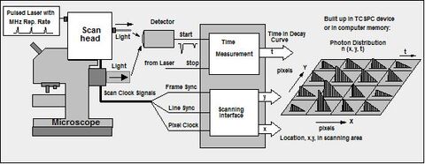
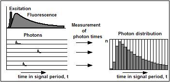
Time-correlated single photon counting (TCSPC) is a common technique to measure fluorescence decays in the time domain. In principle, single photon events are detected and their time of arrival is correlated to the laser pulse, which was used for excitation of the sample. By using a pulsed laser with a high repetition rate, this process can be repeated many times so that a photon distribution over the time and the spatial coordinates is built up. Fig. 1 explains the operation principle in more detail.

Figure 1: Operation principle of time-correlated single photon counting (TCSPC) measurements. The sample is excited by a pulsed laser source with a high repetition rate. Photons emitted by the sample are detected with a high-gain photomiltiplier and the time with respect to the excitation pulse is measured. By counting many events a histogram of the photon distribution over time is built up. (Figure reproduced from: Becker, The bh TCSPC Handbook, 6th ed., 2014, p. 69 - Link)

On the stage of a laser scanning microscope the arrival time of the photon and the coordinates of the laser beam in the scan area are used to build up a 3-dimensional matrix, in which for each pixel of the scanned image not only the fluorescence intensity, but a fluorescence decay curve is stored.

Figure 2: TCSPC setup for fluorescence lifetime imaging (FLIM). The TCSPC module receives the timing pulse, the PMT channel number, and the scan clock signals (frame sync, line sync and pixel clock) from the scanning unit of the microscope. For each photon, the TCSPC module determines the location within the scanning area (x and y) and the time of the photon in the laser pulse sequence (t). These values are used to address a memory in which the detection events are accumulated. Thus, in the memory the distribution of the photon density over x, y and t is built up (Figure reproduced from: Becker, Medical Photonics 27, 41-61 (2015)).

Staff Teaching
How to find us in Jena? Open Positions
Research Contact Information
Methods
Impressum
Datenschutz
FSU
VUD